Amniocentesis
Amniocentesis is a specialized diagnostic test that gently bridges science and the miracle of life, offering a deeper understanding of a baby’s genetic health and development while still within the womb. Usually performed during the middle months of pregnancy, this delicate procedure involves collecting a small sample of amniotic fluid– the clear, protective liquid that lovingly cushions and nourishes the baby inside the uterus.
Within this sacred fluid lie tiny fetal cells and vital biochemical markers that hold profound insights into the baby’s chromosomes, genetic makeup, and overall well-being. When studied in the laboratory, they help doctors identify certain genetic or chromosomal conditions early, allowing time for thoughtful guidance, informed decisions, and specialized care if needed.
When is Amniocentesis done?
- It is performed between 15 and 20 weeks of pregnancy.
- It shows a risk of chromosomal or genetic abnormalities.
- It reveals a family history of genetic or chromosomal disorders.
- It helps doctors assess fetal infection, anemia, or lung maturity later in pregnancy.
How is Amniocentesis done?
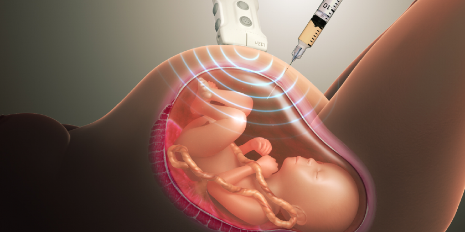
- Ultrasound Guidance: The doctor first performs an ultrasound scan to locate the baby, placenta, and pockets of amniotic fluid, ensuring the safest path for the procedure.
- Cleaning & Preparation: The mother’s abdomen is gently cleaned with an antiseptic solution. Usually, no anesthesia is required, though a local numbing agent may be used for comfort.
- Fluid Collection: A thin, hollow needle is carefully inserted through the abdominal wall into the uterus. A small amount (about 20 ml) of amniotic fluid is withdrawn. The baby remains safe throughout, as the process is monitored in real time with ultrasound.
- Sample Testing: The collected fluid is sent to a laboratory where fetal cells are analyzed for chromosomal, genetic, or metabolic conditions.